
 What sets hooves apart, and makes them more vulnerable than other skin (dermal) structures, is their function. Hooves are critical to overall equine health and can serve as a reliable indicator of your horse’s dermal health.
A horse’s hoof serves to protect its foot. The hoof, and its underlying structures, is a type of integument like the skin on the rest of the body. Although there are differences in the way the hoof appears compared to skin, there are many similarities in both structure and response to injury. The skin and the hoof are both composed of three layers: the epithelium, the dermis, and the subcutaneous tissue, progressing from superficial to deep. The hoof is formed by epithelial keratinization over a greatly modified dermis (sometimes called the corium) that is continuous with the common dermis of the skin at the coronet/coronary band (the junction between the skin and hoof). The subcutaneous tissue of the integument of the foot is also highly specialized.
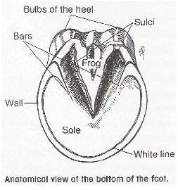
Unlike skin, which tends to be topographically uniform, the integument of the foot is divided topographically into several regions: wall, periople, sole, and frog. These regions reflect the different appearance, structure, and function of the integument in each part of the foot. The wall is the part of the hoof visible in the standing horse. It extends from the coronet to the sole. The wall grows from the coronet and takes about one year to reach the sole. The wall is highest and thickest dorsally (toward the front of the horse) at the toe. It decreases in height and thickness over the sides (quarters) until it is reflected upon itself, forming the rounded heels at the back of the hoof. The inflected parts continue forward for a short distance as the bars that are visible beside the frog when the hoof is raised.
The periople serves as a transition between the soft skin and the hard hoof wall. It starts as a band of soft, rubbery horn a few millimeters thick near the coronet and becomes a thin glossy layer distally (toward the ground). This band widens toward the palmar (toward the back of the horse) aspect where it covers the bulbs of the heels and blends with the base of the frog. The sole fills the space between the wall and the frog and forms most of the underside of the hoof. The sole is slightly concave so that only the distal edge of the wall and the frog make contact on firm ground. The sections between the bars and quarters are called the angles of the sole. The junction between the sole and the wall is the white line. The internal rim of the white line is where a farrier places nails when shoeing.
The wedge shaped frog is a pad of soft horn that projects from behind into the sole between the bars. Its wide base closes the gap between the heels, and the frog’s bulbs of the heels overhang the heels of the wall. The frog has both a central groove and a groove on each side separating it from the bars and the sole.
The specialized layers of the hoof react differently to trauma than the skin does. The hard hoof wall will deflect a minor blow that would otherwise damage skin. Small defects in the outer layers of the hoof are generally of no clinical significance. More serious injury causes cracks that are deflected outward to the exterior surface of the foot instead of toward the dermis. The wall tends to fracture along predetermined planes rather than tear irregularly. The edges of an incision type of wound, for example created by a horse kicking against an edge of sheet metal, will stay together instead of gaping open like skin.
Quarter cracks of the hoof wall are common, typically starting at the coronary band and continuing distally (toward the ground). They are usually full thickness extending into the dermis of the hoof and often lead to instability, inflammation, and infection. The forcible tearing away of (avulsions) of the hoof wall usually involve the wall at the quarters or the heels and can be incomplete or complete. An incomplete hoof wall avulsion remains attached to the foot along at least one margin, while in a complete hoof wall avulsion the affected wall is completely separated from the foot.
Puncture wounds are common injuries to the ground surface of the foot and occasionally occur at the coronary band.
Determining the cause of a foot wound is crucial to correcting the problem and preventing future problems. While injuries can occur at any time due to accidents, some of the causes of foot wounds occupations that put extra stresses on the foot and require frequent shoeing.
- Inherited traits resulting in a poorly shape hoof and/or poor conformation
- Interference of one foot or limb with another foot or limb.
- Hoof wall lesions
Interference injuries are caused when the shoe or foot of one limb strikes another foot or limb resulting in injury.
Causes of interference injuries include:
- Poor conformation
- Exercise surface
- Imbalance between the rider and the horse
- Immature horse
- Unfit horse
- Imbalance due to poor shoeing
- Imbalance caused by hoof overgrowth.
Removing the cause and correctly adjusting foot balance are the only permanent solutions for preventing hoof injuries.